
Brain Vascular Reconstruction, Simulation, and Visualization
October 14th, 2012
Categories: Applications, Software, Virtual Medicine, Visualization, VR
Authors
Marrinan, T., Gould, I., Hsu, C., Linninger, A.About
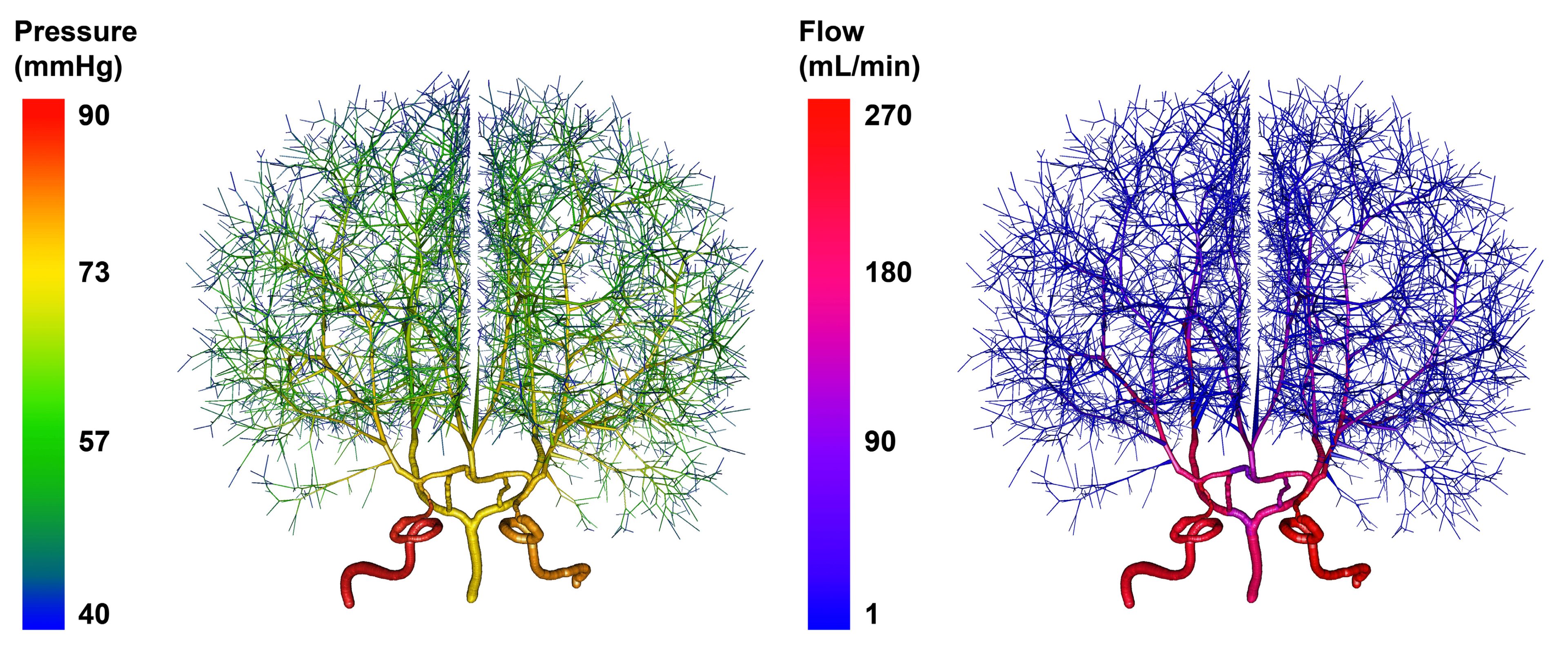
Current techniques in medical imaging and analysis primarily focus on recording information about one specific physiological property at a time. Various modalities such as magnetic resonance, computed tomography, and digital subtraction angiography are each suited towards different tasks. In order to improve surgical planning, physicians would benefit from patient specific computational models built from medical images. These models could be used in order to run simulations and simultaneously gather physiological information that would otherwise require multiple imaging modalities or be impossible to measure with current technology.
We present a pipeline for processing medical data and executing computational simulations to enhance the information conveyed in standard medical imaging. Our work focuses on the whole brain, where we’ve developed tools that allow vasculature to be analyzed in three-dimensions, at high resolutions, and with multiple relevant data sets overlaid on the vascular structure. In order to avoid confusion and misinterpretations, we have the ability to render simulated data such that it mirrors raw medical images and vascular reconstructions.
Keywords: Medical image reconstruction, computer simulation, voxelization, vasculature
Index Terms: I.4.1 [Image Processing and Computer Vision]: Digitization and Image Capture - Imaging Geometry; J.3 [Life and Medical Science]: Health
Resources
Citation
Marrinan, T., Gould, I., Hsu, C., Linninger, A., Brain Vascular Reconstruction, Simulation, and Visualization, Proceedings of VisWeek 2012, Seattle, WA, October 14th, 2012.