
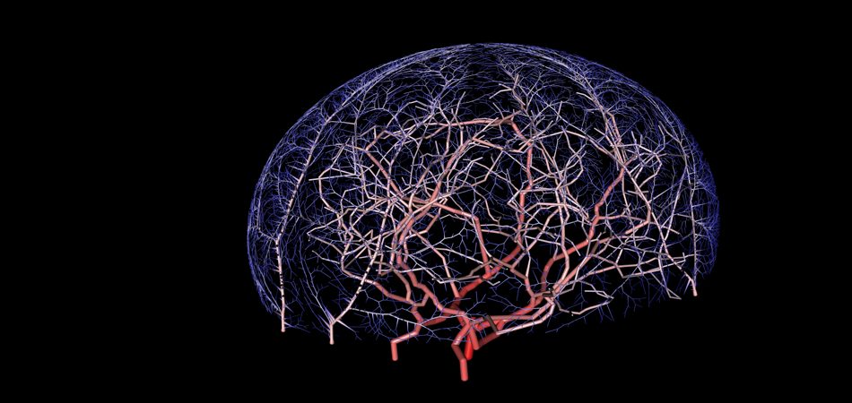
Reconstructing the Cerebral Vascular Network
June 1st, 2011 - May 15th, 2013
Categories: Applications, Education, MS / PhD Thesis, Virtual Medicine, Visualization
About
Researchers from the UIC Departments of Computer Science and BioEngineering propose to develop graphical stereoscopic visual applications for a standard single monitor as well as an ultra-high resolution multi-paneled platform. The visualization will focus on viewing 4D-dimensional (space and time) medical images from angiography, MRI, CT, and computer simulations for an individual patient. The graphical applications developed as part of “Artificially created cortical functional blood unit” will allow neurosurgeons to explore patient-specific data by efficiently organizing and simultaneously displaying all relevant information. Software is being developed to extract quantitative information from patient-specific images such as contrast transit times, as well as sizes and dimensions of anatomic regions of interest. Interfaces are also being created to overlay hemodynamic simulation results into the graphical environment. The visualization tools will enhance insight about the pathology of patients suffering from neurovascular disease, especially stroke. Improved preparation for surgical intervention is expected to empower surgeons to choose better medical treatment paths.
A semi-cylindrical multi-paneled mini-CAVE prototype is being built to render the graphical software applications mentioned above. This virtual reality environment will afford surgeons the ability to incorporate stereo depth perception and immerse themselves fully in a high-resolution environment created by the medical image. User interaction with touch inputs, tablet control, and body motion will provide a novel means of data exploration. This mini-CAVE will offer physicians a means to explore physiologically accurate patient-specific medical images with links to quantitative computer analysis. This research aims to create a three-dimensional virtual reality interface that allows physicians to interact with existing advanced scientific tools in an intuitive fashion. Tools are being created for surgical planning of medical interventions for cerebrovascular diseases that will address spatial constraints such as trajectories through bone and brain tissue, as well as avoidance of white matter fiber tracts. To assess the benefits for 4D surgical planning, a user study is proposed comparing surgeons provided with conventional standard images versus the mini-CAVE exploration environment.