
Mapping of Cognitive Function with Subdural Electrodes & Registration of Cerebral Evoked Potentials on 3-D MRI
August 1st, 1991 - August 1st, 1992
Categories: Applications, Virtual Medicine, Visualization
About
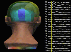
This application is an example of sensory evoked potentials mapped onto a 3D surface model of a human head.
Included in the model is a 3D brain surface originally from MRI data and polygonalized to produce real-time graphics. The head model is a clay model scanned in using a Cyberware laser scanner, and is also polygonalized.
The voltage potentials are measured off the head using 21 separate electrodes. The electrodes measure brain activity. The recordings were made during an experiment in which a patient is shown a checker- board pattern. In addition, recordings were made during a stimulation of the tibial nerve. The resultant voltage potentials were recorded over brief time periods (approximately 10 milliseconds) and the range of voltages is mapped onto a color scale. The color values are then placed on the head model and interpolated producing color gradations. Areas of increased, decreased, and static brain activity can then be visualized.
“Mapping of Cognitive Function with Subdural Electrodes?” premiered at the Showcase exhibit at SIGGRAPH 92, Chicago.